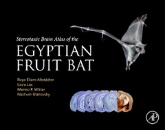
Stereotaxic Brain Atlas of the Egyptian Fruit Bat
Auteurs : Eilam-Altstadter Raya, Las Liora, Witter Menno, Ulanovsky Nachum

The Stereotaxic Brain Atlas of the Egyptian Fruit Bat provides the first stereotaxic atlas of the brain of the Egyptian fruit bat (Rousettus aegyptiacus), an emerging model in neuroscience. This atlas contains coronal brain sections stained with cresyl violet (Nissl), AChE, and Parvalbumin ? all stereotaxically calibrated. It will serve the needs of any neuroscientist who wishes to work with these bats ? allowing to precisely target specific brain areas for electrophysiology, optogenetics, pharmacology, and lesioning. More broadly, this atlas will be useful to all neuroscientists working with bats, as it delineates many brain regions that were not delineated so far in any bat species. Finally, this atlas will provide a useful resource for researchers interested in comparative neuroanatomy of the mammalian brain.
Introduction Methods (Surgery, Histological methods, Stereotaxic reference system and correcting for shrinkage, Delineation of structures) References Index of structures Index of abbreviations Figures: Color plates in stereotaxic coordinates Appendixes: TH staining, ChAT staining
Dr. Las is an Associate Research Scientist at the Weizmann Institute of Science, where she investigates the neural mechanisms of navigation and social cognition in the bat hippocampal formation, along with Nachum Ulanovsky. After completing her Ph.D in auditory neurophysiology, she completed her postdoctoral training with Michale Fee at MIT investigating the mechanisms underlying motor learning in song birds. Her interest lies in understanding the neural mechanisms of natural behaviors, and in developing novel neurotechnologies.
Dr. Witter is a Professor of Neuroscience at the Norwegian University of Science and Technology (NTNU), Norway. He was trained in comparative neuroanatomy and currently studies the functional anatomy of hippocampal-cortical networks with a strong focus on the comparison between neural networks in subdivisions of the entorhinal cortex. He is actively involved in several international consortia on brain atlases and brain nomenclature.
Dr. Ulanovsky is a Professor of Neuroscience at the Weizmann Institute of Science, Israel. He studies the neural basis of navigation and spatial cognition, as well as social cognition, in the mammalian hippocampal formation – using bats as a novel animal model that he pioneered. His group develops wireless-electrophysiology devices, which enabled the discovery of 3D place-cells and 3D head-direction cells in flying bats, goal-vector cells, as well as social place-cells – neurons that represent the positions of other individuals. He seeks to create a "Natural Neuroscience" approach for studying the neural basis of behavior – tapping into the animal's natural behaviors in complex, la
- Provides detailed and accurate stereotaxic coverage of the Egyptian fruit bat forebrain
- Contains 87 plates of coronal sections of adult Egyptian fruit bats, each with one Nissl-stained hemisphere and the other stained either for AChE or Parvalbumin
- Delineates brain structures in the bat brain
- Serves as an essential tool for directing electrophysiology, imaging, optogenetics, pharmacology and lesioning in Egyptian fruit bats, and bats more generally
- Provides a rich resource for comparative neuroanatomy of the mammalian brain
Date de parution : 04-2022
Ouvrage de 162 p.
21.5x27.6 cm


