Myopathies and Tendinopathies of the Diabetic Foot Anatomy, Pathomechanics, and Imaging
Langue : Anglais
Auteur : Pierre-Jerome Claude
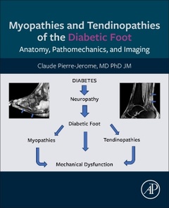
Myopathies and Tendinopathies of the Diabetic Foot is a unique reference of valuable instructive data that re-enforces the understanding of myopathies and tendinopathies related to diabetes-induced Charcot Foot. Diabetic myopathies usually precede other complications (i.e. deformity, ulceration, infection) seen in the diabetic foot. Oftentimes, these myopathies may be isolated especially during their initial stage. In the absence of clinical information relevant to diabetes, the solitaire occurrence of myopathies may lead to confusion, misinterpretation, and misdiagnosis. The misdiagnosis can cause delay of management and consequent high morbidity. This book emphasizes the complications of diabetic myopathies and tendinopathies and all their aspects including pathophysiology, pathomechanics, imaging protocols, radiological manifestations, histological characteristics, and surgical management.Diabetes type II and its complications (diabetic myopathies and tendinopathies) have reached a dreadful high incidence worldwide. Likewise, the need for better understanding of these complications becomes indispensable. In this book, the readers of all genres will find all they need to know about these conditions. The book serves as a classic academic reference for educators, health care specialists, health care givers and health care students.
1. Anatomy of the foot intrinsic muscles
2. Biomechanics of the foot intrinsic muscles
3. Anatomy of the lower leg extrinsic muscles
4. Biomechanics of the lower leg extrinsic muscles
5. Physiology of the skeletal muscles (in general)
6. Mechanism (Pathophysiology) of diabetic foot myopathies
7. Epidemiology (global incidence) of diabetic foot myopathies
8. The Myotendinous Junction: definition, infrastructure
9. Concept of diabetic tendino-myopathy
10. Muscular edema in the diabetic foot Differential diagnosis of muscular edema
11. Muscular atrophy (amyotrophy)in the diabetic foot
12. Muscular infection (pyogenic myositis)
13. Vascular supply to the intrinsic and extrinsic muscles Pathology: muscular infarction / necrosis
14. Innervation of the intrinsic and extrinsic foot muscles Pathology: muscular denervation
15. Imaging of the diabetic myopathies
(1): Radiography and Computed Tomography
16. Imaging of the diabetic foot myopathies
(2): Ultrasonography
17. Imaging of the diabetic foot myopathies
(3): Magnetic Resonance Imaging (MRI)
18. Diabetic foot myopathies: associated soft tissues lesions a (tendinopathies, fasciitis, cellulitis, ulcerations, sinus tract)
19. Diabetic foot myopathies: associated bony lesions (edema, necrosis, infection, fracture, dislocation)
20. Diabetic myopathies: biopsy and pathological evidence
21. Clinical diagnosis and non-surgical management of diabetic myopathies
22. Surgical management of diabetic foot myopathies and tendinopathies
23. COVID-19 and Diabetic Myopathies
Dr. Pierre-Jerome has over 40 years of experience in Radiology and research including diabetes, skeletal muscle and bone marrow. Author of several publications in International Journals. Co-author of five books in Radiology. Reviewer for US and European Journals. Author of two novels. He’s a member of several Medical Associations and fluent in English, French, Spanish and Norwegian.?He was the visiting Associate Professor in University of Rochester, New York and worked as Faculty and Director of International Exchange Program for the Musculoskeletal Division at Emory University School of Medicine, Atlanta GA, USA.?Presently he is working at the Akershus University Hospital in Oslo, Norway.
- Presents pathophysiology, pathomechanics, imaging protocols, radiological manifestations, histological characteristics, and surgical management
- Includes dedicated chapters on tendons and myotendinous junction which are anatomical components frequently ignored in the study of muscles
- Descriptions of diabetic foot myopathies are featured by magnetic resonance imaging (MRI)
- Illustrations of myopathies and tendinopathies with state-of-the-art MRI images and histological images, MR imaging protocols for myopathies
- Anatomical descriptions of all intrinsic and extrinsic muscles
- Detailed information on biomechanics of these muscles
Date de parution : 10-2024
Ouvrage de 350 p.
19x23.3 cm
Thème de Myopathies and Tendinopathies of the Diabetic Foot :
Mots-clés :
Diabetes Mellitus; Plantar muscle; Extrinsic muscle; Lower Extremity; Aponeurosis; Ultrastructure; Pathophysiology; Pathologies; Pedal deformity; Myotendinous Junction; Myositis; Edema; Amyotrophy; Denervation; Ischemia; Necrosis; Pyomyositis; Imaging Modalities; Diagnostic Protocols; Clinical Signs; Biopsy; Pathological Evidence; and Surgical Approach
© 2024 LAVOISIER S.A.S.