Clinical-MRI Correlations of Anterior Knee Pain , 1st ed. 2023 Common and Uncommon Causes
Auteurs : Codorean Ioan I., Codorean Ion Bogdan
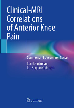
The book addresses comprehensively the normal and pathological MRI appearance of the structures of the anterior compartment of the knee, potential sources of pain, in a systematic way, on anatomical layers, from superficial to deep, respectively, from prepatellar soft tissues to intra-articular structures (the synovial lining, patellar and trochlear cartilage).
Anterior knee pain can affect any age group and causes a non-specific clinical picture, making it difficult to establish a precise diagnosis and proper management. MRI is currently the standard gold investigation and findings of common an uncommon causes of anterior knee pain are presented.
Written by a radiologist and an orthopaedic surgeon with longstanding experience in MRI of musculoskeletal pathology and in sports traumatology, respectively, the book also presents a unique selection of 80 clinical cases of common and less common pathological conditions of anterior knee pain and subdivided in four groups, according to age, from children and adolescents to older adults.
Radiologists and orthopaedic surgeons, as well as sport medicine specialists and physiatrists, will find in this book an invaluable tool for their clinical practice.
Prof. Univ. Dr. Ioan CODOREAN is a primary physician in two medical specialties: Radiology – Medical Imaging and Nuclear Medicine and obtained the title of PhD D in 1998 with the thesis “Correlation Tomodensitometric and Scintigraphic Study of Ischemic Stroke in the Carotid Territory”.
He worked as a military doctor in the Central Military Hospital of Bucharest from 1976 until 2011 when he retired. He continued to work in the Central Military Hospital until 2016 (as a professor at the Carol Davila University of Medicine and Pharmacy.Throughout his medical career, he has contributed to the introduction, promotion, and development of medical imaging techniques in both nuclear medicine and radiology.
In 1977 he established the Nuclear Medicine Laboratory and introduced into clinical practice the techniques of scintigraphic imaging and radioimmunological dosing of hormones and radiochemical techniques for determining the parameters of ferrokinetics
He was President of the Romanian Society of Nuclear Medicine (SRMN) (2000-2005, 2005-2010). Since 2010 he has been an honorary member of this Society.
He was founding member of the Society of Magnetic Resonance in Medicine of Romania (SRMR) in 2006 and of the Romanian working Group of ISMRM “Romanian Chapter of ISMRM” in 2008, and was elected President of the Society of Magnetic Resonance in Medicine of Romania (SRMR) in 2007-2009 and 2009-2011
He has published as author and co-author 4 books, 17 book chapters, and over 150 scientific communications with abstracts in the volume of scientific manifestations and articles in specialized journals.Prof. I. Codorean also organized several courses and congresses:
- Executive President of three National Congresses of Nuclear Medicine with international participation (2001, 2006, 2010)
- Director of the First In
Addressing common and uncommon causes of anterior knee pain with MRI correlation
Featuring over 1000 images and a most interesting selection of clinical cases according to age groups
Presenting MRI of normal and pathological appearance of anterior knee on anatomical layers
Date de parution : 10-2023
Ouvrage de 372 p.
17.8x25.4 cm
Disponible chez l'éditeur (délai d'approvisionnement : 15 jours).
Prix indicatif 210,99 €
Ajouter au panier