FRET - F¿rster Resonance Energy Transfer From Theory to Applications
Coordonnateurs : Medintz Igor L., Hildebrandt Niko
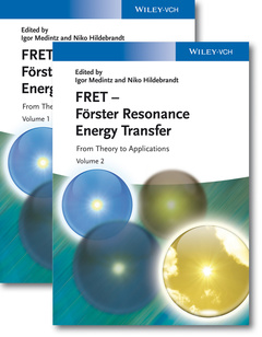
Meeting the need for an up-to-date and detailed primer on all aspects of the topic, this ready reference reflects the incredible expansion in the application of FRET and its derivative techniques over the past decade, especially in the biological sciences. This wide diversity is equally mirrored in the range of expert contributors.
The book itself is clearly subdivided into four major sections. The first provides some background, theory, and key concepts, while the second section focuses on some common FRET techniques and applications, such as in vitro sensing and diagnostics, the determination of protein, peptide and other biological structures, as well as cellular biosensing with genetically encoded fluorescent indicators. The third section looks at recent developments, beginning with the use of fluorescent proteins, followed by a review of FRET usage with semiconductor quantum dots, along with an overview of multistep FRET. The text concludes with a detailed and greatly updated series of supporting tables on FRET pairs and Förster distances, together with some outlook and perspectives on FRET.
Written for both the FRET novice and for the seasoned user, this is a must-have resource for office and laboratory shelves.
Preface xv
List of Contributors xix
Part One Background, Theory, and Concepts 1
1 How I Remember Theodor Forster 3
Herbert Dreeskamp
2 Remembering Robert Clegg and Elizabeth Jares-Erijman and Their Contributions to Fret 9
Thomas M. Jovin
2.1 Biographical Sketch of Bob Clegg 10
2.2 Biographical Sketch of Eli Jares-Erijman 11
2.3 The Pervasive Influence of Gregorio Weber 12
2.4 Contributions by Bob Clegg to Fret 12
2.5 Contributions by Eli Jares-Erijman to FRET 16
2.6 A Final Thought 18
References 19
3 Forster Theory 23
B. Wieb van der Meer
3.1 Introduction 23
3.2 Pre-Förster 23
3.3 Bottom Line 25
3.4 9000-Form, 9-Form, and Practical Expressions of the R06Equation 26
3.5 Overlap Integral 28
3.6 Zones 31
3.7 Transfer Mechanisms 33
3.8 Kappa-Squared Basics 34
3.9 Ideal Dipole Approximation 35
3.10 Resonance as an All-or-Nothing Effect 36
3.11 Details About the All-or-Nothing Approximation of Resonance 39
3.12 Classical Theory Completed 41
3.13 Oscillator Strength–Emission Spectrum Relation for the Donor 42
3.14 Oscillator Strength–Absorption Spectrum Relation for the Acceptor 43
3.15 Quantum Mechanical Theory 44
3.16 Transfer in a Random System 47
3.17 Details for Transfer in a Random System 48
3.18 Concentration Depolarization 51
3.19 Fret Theory 1965–2012 52
References 59
4 Optimizing the Orientation Factor Kappa-Squared for More Accurate Fret Measurements 63
B. Wieb van der Meer, Daniel M. van der Meer, and Steven S. Vogel
4.1 Two-Thirds or Not Two-Thirds? 63
4.2 Relevant Questions 65
4.3 How to Visualize Kappa-Squared? 65
4.4 Kappa-Squared Can Be Measured in At Least One Case 68
4.5 Averaging Regimes 70
4.6 Dynamic Averaging Regime 72
4.7 What Is the Most Probable Value for Kappa-Squared in the Dynamic Regime? 76
4.8 Optimistic, Conservative, and Practical Approaches 83
4.9 Comparison with Experimental Results 85
4.10 Smart Simulations Are Superior 90
4.11 Static Kappa-Squared 92
4.12 Beyond Regimes 101
4.13 Conclusions 102
References 103
5 How to Apply Fret: From Experimental Design to Data Analysis 105
Niko Hildebrandt
5.1 Introduction: Fret – More Than a Four-Letter Word! 105
5.2 Fret: Let’s Get Started! 106
5.3 Fret: The Basic Concept 107
5.4 Fret: Inevitable Mathematics 112
5.4.1 Förster Distance (Or Förster Radius) 112
5.4.2 Fret Efficiency 113
5.4.2.1 Determination by Donor Quenching 113
5.4.2.2 Determination by Acceptor Sensitization 113
5.4.2.3 Determination by Donor Quenching and Acceptor Sensitization 114
5.4.2.4 Determination by Donor Photobleaching 115
5.4.2.5 Determination by Acceptor Photobleaching 115
5.4.3 Fret with Multiple Donors and/or Acceptors 116
5.5 Fret: The Experiment 118
5.5.1 The Donor–Acceptor Fret Pair 118
5.5.2 Förster Distance Determination 119
5.5.3 The Main Fret Experiment 122
5.5.3.1 Steady-State Fret Measurements 123
5.5.3.2 Time-Resolved Fret Measurements 130
5.5.3.3 Interpretation of Time-Resolved FRET Data 133
5.6 Fret beyond Förster 139
5.6.1 Time-Resolved Fret with Lanthanide-Based Donors 140
5.6.1.1 Terbium to Quantum Dot Fret Using Time-Resolved Donor Quenching and Acceptor Sensitization Analysis 141
5.6.2 BRET and CRET 147
5.6.3 Energy Transfer to Metal Nanoparticles (FRET, NSET, DMPET, NPILM, etc.) 148
5.6.4 Other Transfer Mechanisms 150
5.6.4.1 Electron Exchange Energy Transfer (Dexter Transfer) 151
5.6.4.2 Charge Transfer (Marcus Theory) 152
5.6.4.3 Plasmon Coupling 153
5.6.4.4 Singlet Oxygen Diffusion 154
5.7 Summary and Outlook 155
References 156
6 Materials for FRET Analysis: Beyond Traditional Dye–Dye Combinations 165
Kim E. Sapsford, Bridget Wildt, Angela Mariani, Andrew B. Yeatts, and Igor Medintz
6.1 Introduction 165
6.2 Bioconjugation 166
6.3 Organic Materials 171
6.3.1 Ultraviolet, Visible, and Near-Infrared Emitting Dyes 171
6.3.2 Quencher Molecules 173
6.3.3 Environmentally Sensitive Fluorophores 175
6.3.4 Dye-Modified Microspheres/Nanomaterials 179
6.3.5 Dendrimers and Polymer Macromolecules 180
6.3.6 Photochromic Dyes 182
6.3.7 Carbon Nanomaterials 186
6.4 Biological Materials 188
6.4.1 Natural Fluorophores 188
6.4.2 Nonnatural Amino Acids 190
6.4.3 Green Fluorescent Protein and Derivatives 192
6.4.4 Light-Harvesting Proteins 200
6.4.5 DNA-Based Macrostructures/Nanotechnology 201
6.4.6 Enzyme-Generated Bioluminescence 201
6.4.7 Enzyme-Generated Chemiluminescence 209
6.5 Inorganic Materials 211
6.5.1 Luminescent Lanthanide Complexes and Doped Nano-/ Microparticles 212
6.5.2 Luminescent Transition Metal Complexes 217
6.5.3 Noble Metal Nanomaterials (Gold, Silver, and Copper) 219
6.5.4 Silicon-Based Materials 222
6.5.5 Semiconductor Nanocrystals 223
6.6 Multi-Fret Systems 231
6.7 Summary and Outlook 236
References 236
Part Two Common Fret Techniques/Applications 269
7 In Vitro Fret Sensing, Diagnostics, and Personalized Medicine 271
Samantha Spindel, Jessica Granek, and Kim E. Sapsford
7.1 Introduction 271
7.2 Small Organic Molecules and Synthetic Organic Polymers 272
7.3 Carbohydrate–Lipid 273
7.4 The Biotin–Avidin Interaction 273
7.5 Proteins and Peptides 275
7.5.1 Binding Proteins 275
7.5.2 Antigens and Epitope-Based Peptide Sequences 277
7.5.3 Peptide Sequences for Enzymatic Sensing 279
7.6 Antibodies 282
7.7 Nucleic Acid (DNA/RNA) 287
7.7.1 Molecular Beacons 288
7.7.2 Polymerase Chain Reaction and Fret 289
7.7.2.1 Fret Hybridization Probes 290
7.7.2.2 TaqMan 291
7.7.2.3 Scorpion Assay 292
7.7.2.4 Others 294
7.7.3 Isothermal Amplification Reactions and Fret 294
7.7.4 Clinical Applications of Nucleic Acid Detection Using Fret 295
7.7.4.1 Detection of Pathogens 295
7.7.4.2 Prognostic and Diagnostic Applications 296
7.7.4.3 Pharmacogenomics and Personalized Medicine 298
7.8 Aptamers 299
7.9 High-Throughput and Point-of-Care Devices 302
7.9.1 PoC Technology Advances 302
7.9.2 PoC Material Advances 304
7.10 Conclusions 305
References 305
8 Single-Molecule Applications 323
Thomas Pons
8.1 Introduction 323
8.2 Single-Molecule Fret of Immobilized Molecules 324
8.2.1 Experimental Setup 324
8.2.1.1 Molecule Immobilization 324
8.2.1.2 Fluorophore Photostability 325
8.2.1.3 Optical Setup 326
8.2.2 Data Analysis 326
8.2.3 Applications 329
8.2.4 Analyzing Complex Fret Trajectories 334
8.3 Single-Molecule Fret of Freely Diffusing Molecules 336
8.3.1 Experimental Setup 336
8.3.2 Applications 337
8.3.3 Advanced Solution smfret Methods 343
8.3.3.1 Alternate Laser Excitation 343
8.3.3.2 Multiparameter Fluorescence Detection 344
8.4 Single-Molecule Fret Studies Involving Multiple Fret Partners 346
8.4.1 Multistep Fret 347
8.4.2 Multi-Acceptor and Multi-Donor Systems 348
8.5 Conclusions and Perspectives 351
References 353
9 Implementation of Fret Technologies for Studying the Folding and Conformational Changes in Biological Structures 357
Philip J. Robinson and Cheryl A. Woolhead
9.1 Introduction to Using Fret in Biological Systems 357
9.2 Förster Formalism in the Determination of Biological Structures 358
9.3 Fret Experiments in Complex Biological Systems 360
9.3.1 The Importance of Experimental Design 360
9.3.2 Site-Specific Labeling and Choosing the Most Effective Fret Pair 361
9.4 Biological Model System 1: The Ribosome 362
9.4.1 Intersubunit Rotation within the Ribosome 363
9.4.2 Dynamic Intrasubunit Movement Within the Ribosome 365
9.5 Biological System 2: Nascent Polypeptide Structure 365
9.6 Biological System 3: Chaperone-Mediated Protein Folding 368
9.6.1 Signal Recognition Particle 368
9.6.2 Trigger Factor 369
9.7 Biological System 4: Mature Protein Folding Intermediates 371
9.7.1 Unfolding Kinetics of Monellin 372
9.7.2 Intermediate Folding State of the Src Homology 3 Domain 374
9.8 Biological System 5: Intersubunit Distance in Multimeric Protein Complexes 375
9.9 Biological System 6: Protein–Protein Interactions in the Assembly of Protein Polymers 378
9.9.1 FtsZ Assembly and Subunit Exchange 379
9.9.2 Defining the Molecular Link in Serpin Polymers 380
9.10 Biological System 7: Fret in Nucleic Acid Systems 385
9.10.1 Determining the Structure and Configuration of DNA Junctions 386
9.10.2 Measuring the Opening and Closing of a Nanoscale DNA Box 388
9.10.3 Fret Between a DNA Polymerase and Its Substrate 390
References 392
10 Fret-Based Cellular Sensing with Genetically Encoded Fluorescent Indicators 397
Jonathan C. Claussen, Niko Hildebrandt, and Igor Medintz
10.1 Introduction 397
10.2 Enzymes 399
10.2.1 Kinase Activity/Phosphorylation 399
10.2.2 Protease Activity 403
10.3 Metabolites 407
10.3.1 Sugars 407
10.3.2 Glutamate 410
10.4 Second Messengers 412
10.4.1 cAMP 412
10.4.2 cGMP 415
10.4.3 Nitric Oxide 417
10.4.4 Calcium 419
10.5 Conclusions 421
References 423
Part Three Fret with Recently Developed Materials 431
11 Fret with Fluorescent Proteins 433
Hiofan Hoi, Yidan Ding, and Robert E. Campbell
11.1 Introduction to FPs 433
11.1.1 Wild-Type FPs 433
11.1.1.1 Natural Sources 433
11.1.1.2 Structure 434
11.1.1.3 Chromophore Formation 436
11.1.2 Engineered FPs for Fret Applications 438
11.1.2.1 Overview 438
11.1.2.2 Blue–Green Fret Pairs 440
11.1.2.3 Cyan–Yellow Fret Pairs 441
11.1.2.4 Fret with Orange, Red, and Far-Red FPs 443
11.1.2.5 Atypical FPs Useful for Fret Applications 445
11.1.3 Why Use FPs for Fret? 446
11.2 Using FPs for Fret Imaging 446
11.2.1 Photophysical Properties and Typical Förster Radii 446
11.2.1.1 Overview 446
11.2.1.2 Spectral Overlap 447
11.2.1.3 Orientation Factors 449
11.2.2 Potential Sources of Artifacts During Fret Imaging 450
11.2.2.1 Photobleaching 450
11.2.2.2 Photoconversion 451
11.2.2.3 pH Dependence 452
11.2.3 Biochemical and Structural Considerations 453
11.2.3.1 General Considerations when Labeling Proteins with FPs 453
11.2.3.2 Labeling Proteins for Intermolecular Fret Experiments 454
11.2.3.3 Labeling Proteins for Intramolecular Fret Experiments 454
11.2.3.4 FP Oligomerization and Fret Efficiency 455
11.2.4 Applications and Examples 458
11.2.4.1 Overview 458
11.2.4.2 Fret Biosensor Case Study 459
11.2.4.3 Fret between FPs and Other Donor or Acceptor Materials 460
11.3 Conclusions 462
References 463
12 Semiconductor Quantum Dots and Fret 475
W. Russ Algar, Melissa Massey, and Ulrich J. Krull
12.1 Introduction 475
12.2 A Quick Review of Fret 476
12.3 Quantum Dots 477
12.3.1 A Brief History 478
12.3.2 The Structure of Quantum Dots: The Core 478
12.3.3 The Optical Properties of Quantum Dots 480
12.3.4 Overcoming the Limitations of Molecular Fluorophores 482
12.3.5 The Structure of Quantum Dots: The Shell 483
12.3.6 Quantum Confinement 485
12.3.7 Quantum Dot Photophysics 488
12.3.8 Quantum Dot Synthesis 491
12.3.9 Quantum Dot Coatings 493
12.3.10 Quantum Dot Bioconjugation 496
12.3.11 Quantum Dot Nomenclature in This Chapter 499
12.4 Quantum Dots and FRET 499
12.4.1 Quantum Dots as Donors 499
12.4.2 Applicability of the Förster Formalism 502
12.4.3 QDs as Acceptors 504
12.4.4 The Importance of Bioconjugate Chemistry 506
12.5 Quantum Dots as Donors in Biological Applications 508
12.5.1 Association and Dissociation to Modulate QD-FRET 508
12.5.1.1 Bioanalysis of Carbohydrates 509
12.5.1.2 Homogeneous Immunoassays 510
12.5.1.3 Hybridization Assays 511
12.5.1.4 Bioanalyses Using Aptamers and DNAzymes 516
12.5.1.5 Bioanalysis of Hydrolytic Enzymes 519
12.5.1.6 Gene Delivery 524
12.5.2 Changes in Distance to Modulate QD-FRET 524
12.5.3 Conformational Insights from QD-FRET 528
12.5.4 Dynamic Modulation of the Spectral Overlap Integral and QD-FRET 530
12.5.5 Single-Pair QD-FRET 534
12.5.6 Solid-Phase QD-FRET 540
12.5.6.1 Biomolecular Surface Tethers 542
12.5.6.2 Chemical Conjugation to an Interface 544
12.5.6.3 Interfacial Ligand Exchange 545
12.5.6.4 Electrostatic Immobilization 547
12.5.6.5 Advantages of Immobilized QDs 548
12.5.7 Photodynamic Therapy 549
12.6 Quantum Dots as Acceptors in Biological Applications 552
12.6.1 Chemiluminescence Resonance Energy Transfer (CRET) 553
12.6.2 Bioluminescence Resonance Energy Transfer (BRET) 555
12.6.3 Lanthanide Donors 560
12.6.4 Quantum Dot Donors (for Quantum Dot Acceptors) 565
12.7 Energy Transfer between Quantum Dots and Other Nanomaterials 569
12.7.1 Gold Nanoparticles 569
12.7.2 Carbon Nanomaterials 575
12.7.2.1 Graphene and Graphene Oxide 575
12.7.2.2 Carbon Nanotubes 577
12.8 Nonbiological Applications of Quantum Dots and Fret 578
12.8.1 Photovoltaic Cells 580
12.8.2 Light-Emitting Diodes (LEDs) 582
12.9 Summary 583
References 584
13 Multistep Fret and Nanotechnology 607
Bo Albinsson and Jonas K. Hannestad
13.1 Introduction 607
13.2 Fundamentals of Multistep FRET 608
13.2.1 Hetero-Fret 609
13.2.2 Multicolor Fret and Alternating-Laser Excitation 611
13.2.3 Homo-Fret 612
13.3 Energy Transfer in Photosynthesis 615
13.4 Photonic Wires and Multistep Fret in Nanotechnology 617
13.4.1 Photonic Wires 617
13.4.2 Beyond Wires 628
13.4.3 Light Harvesting 632
13.4.4 Functional Control 638
13.4.5 Quantum Dots in Multistep Fret 641
13.4.6 Potential Outputs and Uses for Channeled Excitation Energy 643
13.5 Summary 647
13.6 Note Added in Proof 648
References 648
Part Four Supporting Information and Conclusions 655
14 Data 657
Alice G. Byrne, Matthew M. Byrne, George Coker III, Kelly B. Gemmill,Christopher Spillmann, Igor edintz, Seth L. Sloan,and B. Wieb van der Meer
14.1 Tables before 1987 658
14.2 Introduction to the Table of Traditional Chromophores 658
14.3 Forster Distances and Other Fret Data before 1994 703
14.4 Forster Distances for Traditional Probes More Recent Than 1993 703
14.5 Fret Data on Fluorescent Proteins 703
14.6 Fret Data on Quantum Dots 742
14.7 Donor–Acceptor Pairs with a Forster Distance in a Given Range 742
14.8 Table–Reference Directory 744
References 745
15 Outlook on Fret: The Future of Resonance Energy Transfer 757
15.1 A Rosy Crystal Ball View of Fret 757
Thomas M. Jovin
15.2 Do Not Ask What Fret Can Do for You, Ask What You Can Do for Fret 757
B. Wieb van der Meer
15.3 Fret: Future Research with an Exciting Technology 758
Niko Hildebrandt
15.4 Future of Fret 760
Kim E. Sapsford
15.5 Outlook on Single-Molecule Fret 760
Thomas Pons
15.6 Outlook on Fret with fluorescent proteins 761
Robert E. Campbell
15.7 Luminescent Nanoparticles: Scaffolds for Assembling “Smarter” FretProbes 762
W. Russ Algar
References 764
Index 767
Dr. Igor L. Medintz obtained his B.S. and M.S in Forensic Science, followed by a Ph.D. degree in Molecular Biology in 1998 at the City University of New York. He carried out research as a postdoctoral fellow at the University of California Berkeley as well as at U.S. Naval Research Laboratory (NRL) in Washington, D.C. Since 2004, he has been a Research Biologist at NRL where he focuses on developing chemistries to interface nanomaterials with biology and understanding how nanoparticles engage in different types of energy transfer.
Professor Niko Hildebrandt obtained a Diploma in Medical Physics in 2001 at the University of Applied Sciences Berlin and a Ph.D. degree in Physical Chemistry in 2007 at the University of Potsdam, where he also carried out postdoctoral research until 2008. From 2008 to 2010 he was head of the group NanoPolyPhotonics at the Fraunhofer Institute for Applied Polymer Research in Potsdam. Since 2010 he has been Full Professor at Université Paris-Sud, where he is leading the group of NanoBioPhotonics (www.nbp.ief.u-psud.fr) at the Institut d’Electronique Fondamentale with a research focus on time-resolved FRET spectroscopy and imaging for multiplexed nanobiosensing.
Date de parution : 12-2013
Ouvrage de 816 p.
17.5x25.2 cm
Disponible chez l'éditeur (délai d'approvisionnement : 14 jours).
Prix indicatif 244,71 €
Ajouter au panier